Button control
Designed to deliver smooth, controlled fluid extraction with minimal force.

DISCOVER OCUTAP
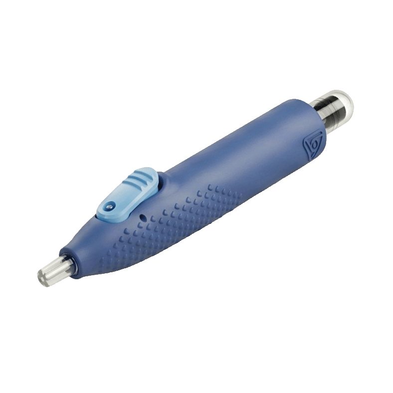
A closed-system, single-use device designed for anterior chamber paracentesis in uveitis and other anterior segment diagnostics, with a detachable vial that ships directly to the lab.
THE CLINICAL NEED
A large, recurring population needs reliable anterior-chamber sampling.
Faster, better-characterized diagnosis can protect sight.
Molecular sampling helps identify the cause, not just the symptom.
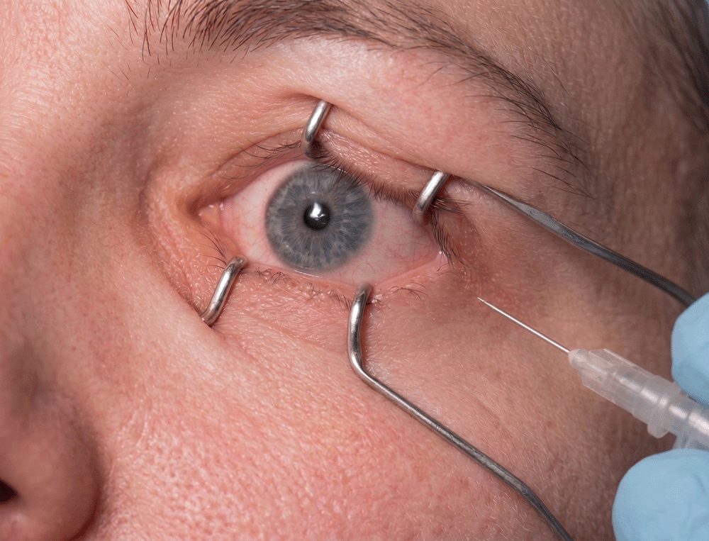
Anterior chamber paracentesis is a high-skill, two-handed procedure performed with a standard syringe and a long needle. The handling is awkward, the sample yield is variable, and a meaningful portion of what is collected is lost during transfer for shipping.
The standard syringe introduces measurable friction across every procedural phase. In surveys of retina, uveitis, glaucoma, and anterior-segment specialists, the same pain points recur, and each carries a number.
Percentages reflect the share of surveyed clinicians reporting each issue.
One-handed control remains elusive with a standard syringe. Stabilizing the eye while drawing fluid demands two hands and steady nerves.
Not all labs accept samples collected in traditional syringes, delaying diagnosis and forcing device compromises.
Needle-stick exposure and injury to adjacent tissue remain persistent concerns during collection and transfer.
Wound leakage after the tap adds risk and follow-up burden.
Monitoring the needle often requires added magnification for control.
A long needle increases the risk of injury during the procedure.
Aqueous molecular biomarkers are moving from research into practice, and reliable ocular fluid sampling is the bottleneck OcuTap is built to remove.
THE SOLUTION
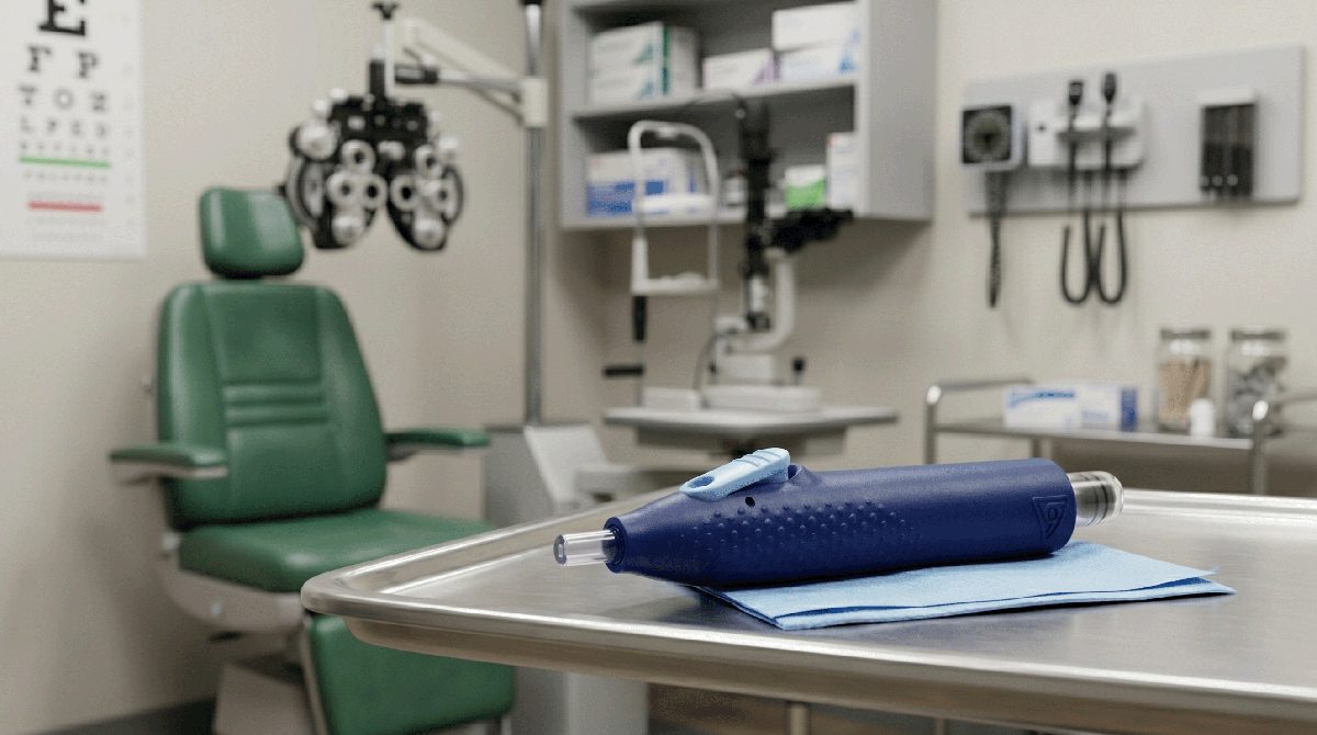
The OcuTap is a single-use paracentesis device designed to simplify aqueous humor extraction with a detachable vial that ships directly to the lab, eliminating manual sample transfers.
Designed to deliver smooth, controlled fluid extraction with minimal force.
A factory-set mechanism is engaged before patient contact, intended to remove the operator’s draw speed as a variable.
Designed for compatibility with standard needles, for quick and secure attachment.
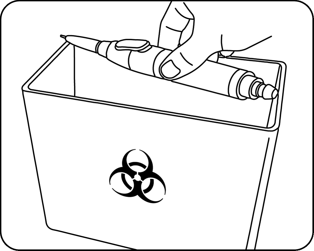
Designed for straightforward disposal — insert the device into a sharps container after vial removal.
Designed to support stability during precise micro-maneuvers.
This clear, sterile sample tube is designed for broad compatibility with diagnostic laboratories.
Watch the video demonstration of the OcuTap procedure or follow the detailed, step-by-step instructions.
The following protocol outlines the intended engineering functionality of the OcuTap device. These instructions are provided for informational and investigational purposes only, pending official regulatory clearance.
The OcuTap has been carefully crafted to improve comfort, control, and efficiency for liquid biopsy of ocular fluid.
First, the device is primed by pulling back the collection tube to an extended position. A click will sound to lock it into place. A needle is attached.
The device is inserted into the eye at the limbus following standard procedures for anterior chamber paracentesis. Collection of the fluid is started with depression of the central button. This opens the valve and allows for steady, smooth, and reliable fluid collection. The users can release the button to stop the fluid if needed.
The system’s design prevents fluid or pressure spikes during collection, and the one-handed operation improves physician control during the procedure, which improves safety for the patient and limits complications.
When the procedure is complete, the button is released and the device is safely withdrawn from the eye. A final push of the button clears the fluid from the internal pathway of the device into the collection tube.
The procedure is complete when the collection tube is twisted to remove it from the device. The device is discarded safely in the sharps container, and a cap is placed on the collection tube for shipping.
Securely attach a sterile needle (not included) to the device. Do not remove the needle cap at this time.
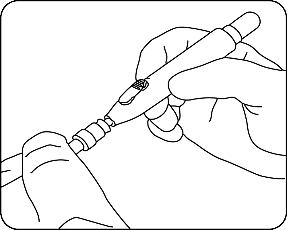
Pull back the collection tube until it reaches an extended position and clicks into place.
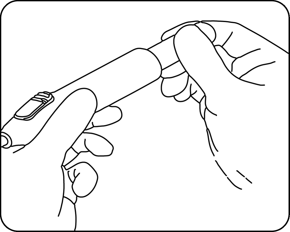
Carefully remove the protective cap from the needle, ensuring the tip remains sterile and does not come into contact with any surrounding surfaces prior to insertion.
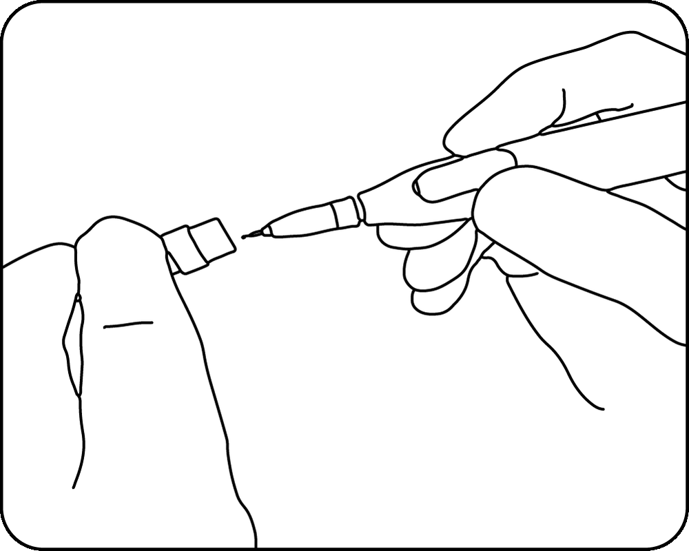
Insert the device into the eye at the limbus, strictly following standard anterior chamber paracentesis procedures. Once safely positioned, depress the central button to open the valve and begin the collection of fluid. If necessary at any point, release the button to immediately stop the fluid flow.
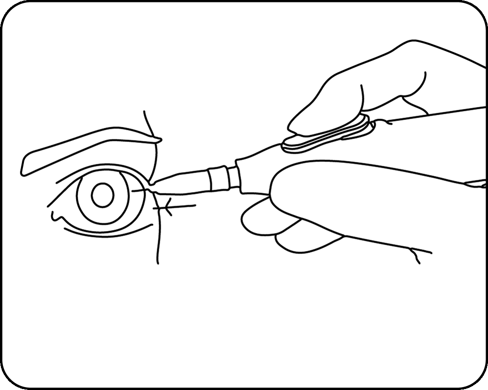
Once the collection is finished, release the button and carefully withdraw the device from the eye.
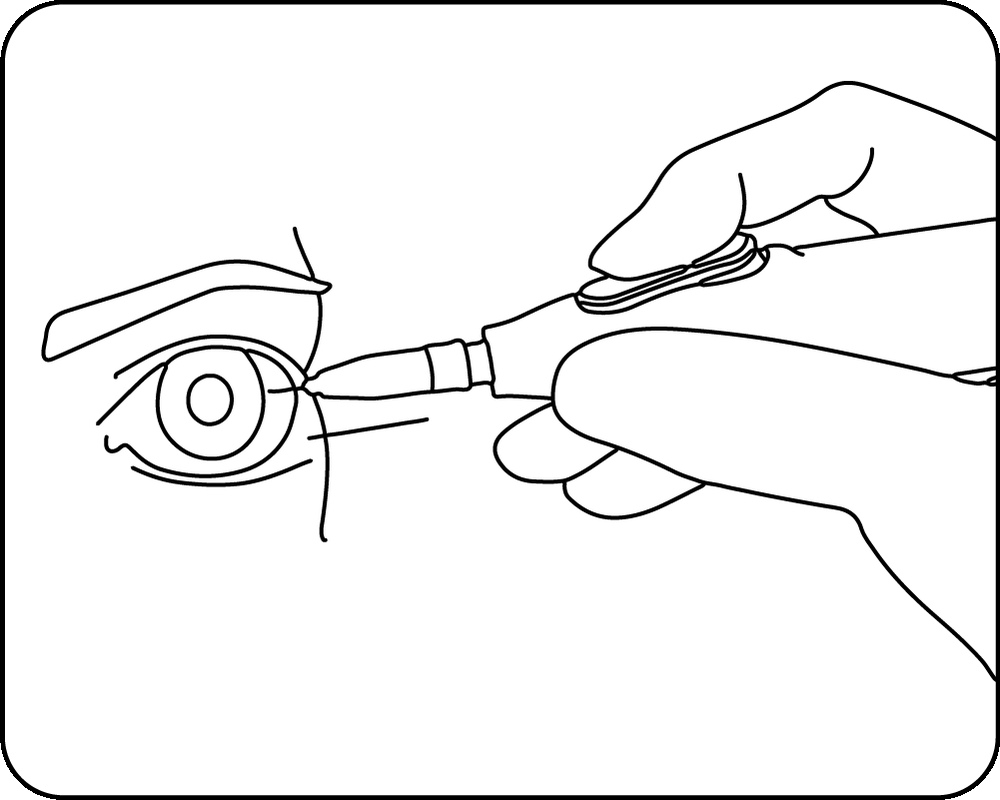
Give the button a final push to move any remaining fluid from the internal pathway into the collection tube.
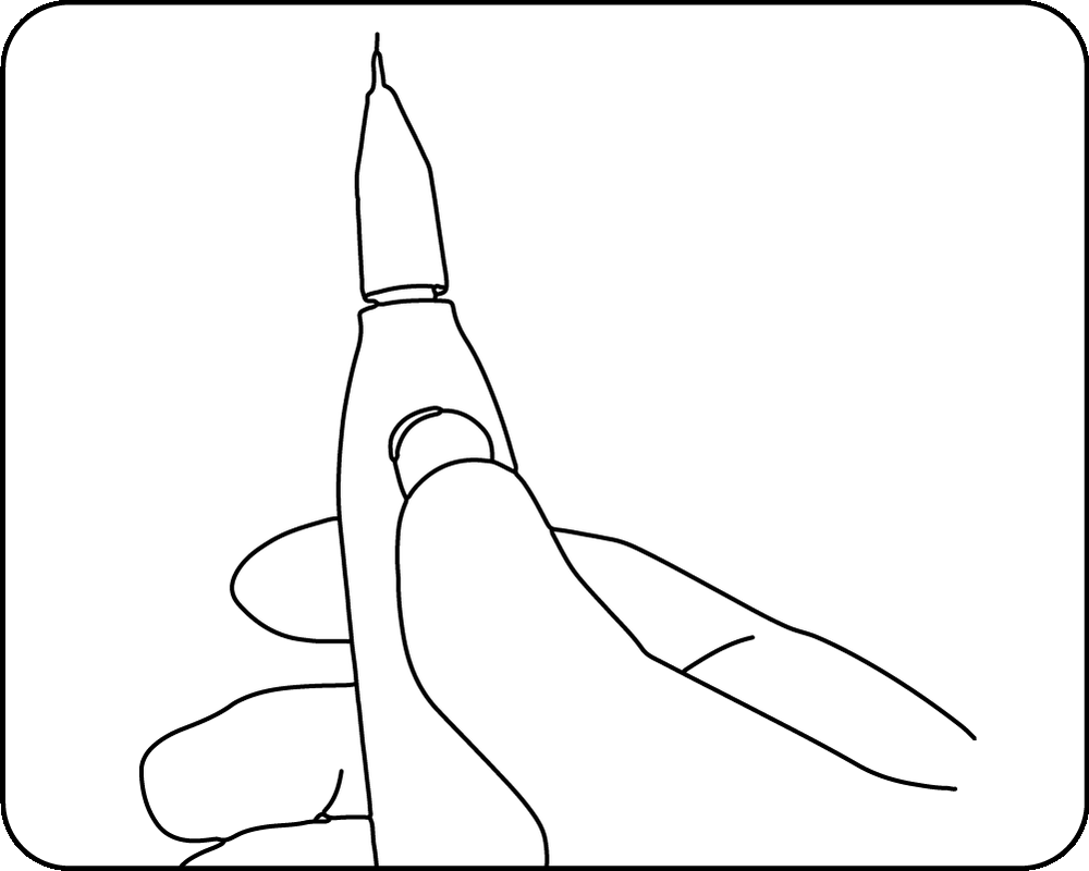
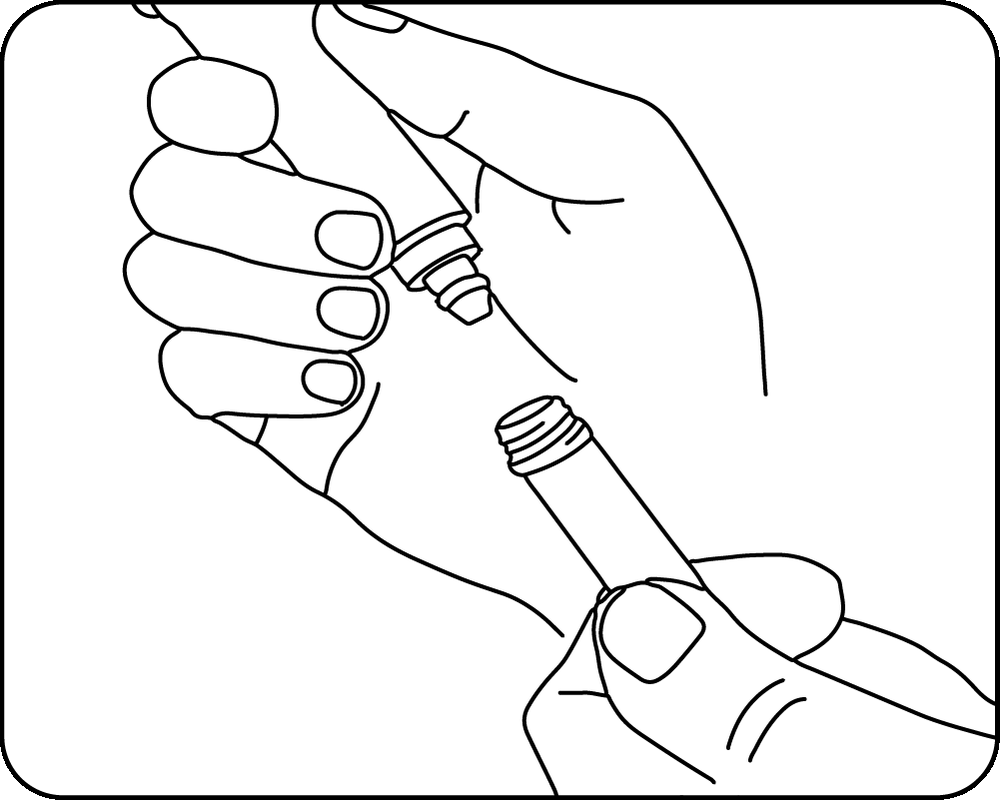
Twist the collection tube to detach and remove it from the primary device.
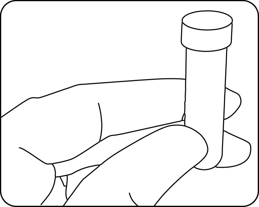
Cap the barrel, and safely discard the syringe body in a sharps container. Place a cap securely on the collection tube for shipping and analysis.
PRELIMINARY INTERNAL DATA
Findings below are from an internal assessment of anterior chamber procedures. Not independently validated by a third party. Data on file.
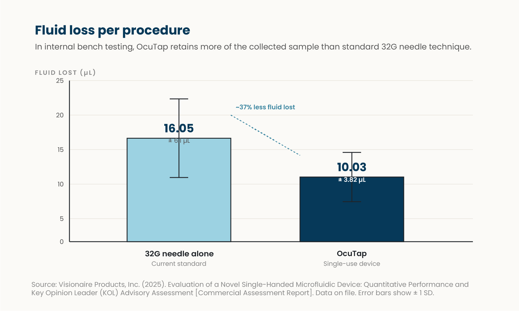
Compared to standard 32-gauge needle technique (10.03 µL vs. 16.05 µL).
In internal testing, OcuTap delivered an average draw rate of approximately 40 µL per second, independent of operator technique.
In an early pilot study, most participating clinicians reported a preference for OcuTap over the standard of care for control and safety. Preliminary data; not independently validated.
OcuTap was developed by a practicing retina specialist and refined with feedback from clinicians who tried it during early development. Here’s what they shared:

“The actuation is controlled and measured, unlike a syringe that can pull fluid too fast, a real safety concern.”

“Not a microliter is wasted, nothing stays behind in the chamber.”

“The device steadies your hand and lets you draw fluid without fumbling, that’s the real advantage.”
CAUTION—Investigational device. Limited by Federal law to investigational use. FDA 510(k) clearance anticipated by the end of 2026.
SAFETY & REGULATORY
FDA review underway for market clearance
Classified under 21 CFR (Code of Federal Regulations) 880.5570 with product code QYL. Standard regulatory pathway for single-use ophthalmic aspiration devices.
CAUTION—OcuTap is investigational and limited by Federal law to investigational use. Regulatory submission is progressing through standard FDA pathways.
We are targeting FDA 510(k) clearance by the end of 2026.
OcuTap is indicated for collection of aqueous humor samples from the anterior chamber in patients with anterior uveitis, infectious keratitis, endophthalmitis, or other anterior segment inflammatory or infectious conditions requiring diagnosis.
OcuTap is contraindicated in patients with corneal scarring or opacity that prevents adequate visualization of the anterior chamber. Use is also contraindicated in patients with shallow anterior chambers where safe needle placement cannot be assured.
Risk of corneal abrasion, hyphema, or lens trauma exists with improper needle placement. Infection or endophthalmitis may occur despite sterile technique. Hypotony or anterior chamber collapse can result from excessive fluid withdrawal.
Maintain strict aseptic technique throughout the procedure. Verify device integrity and sterility before use. Monitor intraocular pressure and anterior chamber stability post-procedure. Counsel patients on post-procedure care and signs of complication.
Disclaimer: Visionaire Products is responsible only for the performance of its devices as intended for specimen collection. Following successful specimen collection and transfer, all aspects of specimen handling, storage, transportation, laboratory testing, analysis, reporting, and disposal are the responsibility of the user and/or receiving laboratory. Visionaire Products assumes no liability for events occurring after specimen collection.
RESOURCES
Instructions for use, a clinical snapshot, and detailed device specifications available on request.
Complete step-by-step guidance for safe device preparation and operation.
A concise overview of device design, performance data and how it fits your clinical workflow.
Detailed engineering and performance specifications. Contains proprietary information and is shared by request rather than published. Request permission and we'll add your email with viewing permission, then send you the link.
OcuTap is pending FDA clearance, anticipated by the end of 2026. Sign up to get updates and we’ll email you when there’s meaningful news to share.